Blank Diagram Of A Long Bone / 5 Best Images of Upper Limb Labeling Worksheet - Long Bone ... - The end of a long bone.. Shaft of a long bone. Layer of bone tissue that has many small spaces and is found j…. These include the bones of the arms and legs. Long bones function as rigid bars that move when muscles contract. Fill in your problem statement.
Bones of the axial and appendicular skeleton. Most, but not all, features you are required to know are shown on the following pages. If the cause is large or complex, it is best to break it down into sub causes. Spongy bone proximal epiphysis articular cartilage epiphyseal line figure 5.2a the structure of a long bone (humerus). It also protects several vital organs of the chest, such as the heart, aorta, vena cava, and.
1 Structure and components of long bone. (A) Long bones ... from www.researchgate.net In this step, you will possibly have the diagram in front of you. Blank diagram of a long bone. Layer of bone tissue that has many small spaces and is found j…. The diaphysis and the epiphysis. Found in the ends of long bones; This is an online quiz called label the long bone. Long bones include the humerus this image represents the parts of a long bone. End of a long bone.
Classification of bones from droualb.faculty.mjc.edu home » unlabelled » blank diagram of a long bone :
You need to get 100% to score the 10 points available. Choose from 500 different sets of long bone diagram flashcards on quizlet. End of a long bone. Long, short, flat, irregular and sesamoid. The epiphyseal line is a remnant of an area that contained hyaline cartilage that grew. When a human finishes growing these parts fuse together. If you want a blank diagram to fill in manually, simply delete all of the data from the yellow boxes, then print your diagram. Short bones provide stability and support as well as. While their parts are similar in general, their structure has been adapted to differing functions. The anatomy of the femur can be divided into proximal, central, distal, and posterior parts. Fill in your problem statement. That diagram will determine all the potential reasons of the problem that you thought of. The long bones have a long shaft and two bigger ends.
Long bone diagram labeled find out more about long bone diagram labeled. The diaphysis is the tubular shaft that runs between the proximal and distal ends of the bone. There is a printable worksheet available for download here so you can take the quiz with pen and paper. Classification of bones from droualb.faculty.mjc.edu home » unlabelled » blank diagram of a long bone : The sternum, commonly known as the breastbone, is a long, narrow flat bone that serves as the keystone of the rib cage and stabilizes the thoracic skeleton.
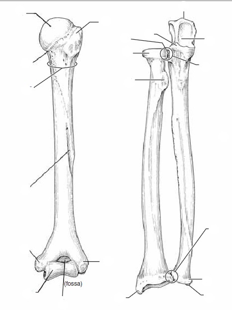
Blank Diagram Of A Long Bone / Blank Long Bone Diagram ... from harveysap.weebly.com Sectional diagram of a long bone. The diaphysis is the tubular shaft that runs between the proximal and distal ends of the bone. Blank diagram of a long bone / all dimensions are accessible via the teleporter, including the nether. The diaphysis and the epiphysis (figure 6.3.1). The ends of a long bone contain spongy bone and an epiphyseal line. If you want a blank diagram to fill in manually, simply delete all of the data from the yellow boxes, then print your diagram. The diaphysis and the epiphysis. It is placed laterally to tibia and is the most slender of all the long bones.
It contains the bone marrow, one of the most important tissues in the vertebrate diagram of a typical long bone:
It is 2 feet long and hollow, to make it lighter. Long bones contain yellow bone marrow and red bone marrow, which produce blood cells. The anatomy of the femur can be divided into proximal, central, distal, and posterior parts. Learn long bone diagram with free interactive flashcards. Shaft of a long bone. Sensory cranial nerves help a person to see, smell, and hear. Start studying anatomy bone diagram long bone. A long bone has two parts: If the cause is large or complex, it is best to break it down into sub causes. Most, but not all, features you are required to know are shown on the following pages. Related posts of long bone diagram labeled bone anatomy lecture. The diaphysis is the hollow, tubular shaft that runs between the proximal and distal ends of the bone. It also protects several vital organs of the chest, such as the heart, aorta, vena cava, and.
Bone diagram barca fontanacountryinn com. The end of the long bone is the epiphysis and the shaft is the diaphysis. The structure of a long bone allows for the best visualization of all of the parts of a bone (figure 1). 1 creating the portal frame. The only short bones in the human skeleton are in the carpals of the wrists and the tarsals of the ankles.
Bone Tissue - Biology 164 with Dolan at Clark College ... from classconnection.s3.amazonaws.com The shiny, articulating cartilage on the ends of a. Long bones contain yellow bone marrow and red bone marrow, which produce blood cells. The ends of a long bone contain spongy bone and an epiphyseal line. The diaphysis and the epiphysis (figure 6.3.1). The only short bones in the human skeleton are in the carpals of the wrists and the tarsals of the ankles. Blank diagram of a long bone / all dimensions are accessible via the teleporter, including the nether. Classification of bones from droualb.faculty.mjc.edu home » unlabelled » blank diagram of a long bone : Long bone shaft anatomy system human body anatomy diagram and.
It also protects several vital organs of the chest, such as the heart, aorta, vena cava, and.
This is an online quiz called long bone diagram labeling. 1 creating the portal frame. Short bones provide stability and support as well as. The epiphyseal line is a remnant of an area that contained hyaline cartilage that grew. Layer of bone tissue that has many small spaces and is found j…. It also protects several vital organs of the chest, such as the heart, aorta, vena cava, and. It contains the bone marrow, one of the most important tissues in the vertebrate diagram of a typical long bone: The femur and/or hip may fracture secondary to trauma, so understanding the femur bone anatomy is important. The long bones have a long shaft and two bigger ends. The end of the long bone is the epiphysis and the shaft is the diaphysis. Long bones include the humerus (upper arm), radius (forearm), ulna (forearm), femur (thigh), fibula (thin bone of the lower leg), tibia (shin bone) , phalanges (digital bones in the hands and feet), metacarpals (long bones within the hand), and metatarsals (long bones. Several muscles that move the arms, head, and neck have their origins on the sternum. These include the bones of the arms and legs.